Saturday, October 19, 2013

WILTIMS #53: Brains and lightsabers

Yesterday was a dramatic day that concluded months of waiting on a couple fronts.

First up was learning how to perform an HEENT (head, eye, ear, nose, throat) exam, which meant finally getting to use the most expensive single item we've had to buy for medical school: our ophthalmoscope/otoscope (eye/ear scope). The cost was particularly annoying because it's not obvious why we need to buy one of these devices in the first place, as they are provided in every exam room and hospital unit in the country. Now, we didn't need to buy the fanciest model, but it was part of a bundle with our blood pressure cuff, reflex hammer, tuning forks, etc, so I splurged. Anyways, it was nice to finally get to use some of this doctor-y stuff!

To examine the eye with an ophthalmoscope, you start out about a foot away from the patient and place your thumb above their eye (to reassure them that you won't smack them in the head with the scope as you move in). You then align the beam of light with the pupil by looking for the “red reflex” which is weirdly named as it is merely the light reflecting off the retina on the back of the eye. This same phenomenon causes red eye in photographs. After finding the red reflex you move closer to the eye, being sure to keep the light centered on the pupil. Once you get close enough, you can see the blood vessels in the back of the eye. In order to see the vessels clearly, you have to counteract the lens of the eye by focusing the lens in the scope. Interestingly, people with different glasses prescriptions will need a different focus setting on the ophthalmoscope.

In anatomy lab yesterday, we finally reached the apex of the class: cutting open the skull and removing the brain. The day before, we had removed the scalp, so we started by placing a rubber band around widest circumference of the skull to mark out our planned cut. We then took an electric oscillating circular saw and cut through the bulk of the skull, trying to leave a small part of the inner surface intact to protect the brain. Oscillating saws, rather than rotary saws are used to cut through bone and casts because they have a much harder time ripping through soft tissue. Next we used a rubber mallet and chisel to break the remainder of the bone and finally expose the brain.

One cool thing about the interior of the skull is that it has indentations in the bone from the blood vessels of the dura mater (outermost meninx). These meningeal layers are the last thing we had to remove to see the brain, which looked... exactly like you'd expect a brain to look. It felt rather rubbery and stiff as a result of the preservation process.

Literally 5 minutes after I was touching the brain of a dead person amidst the smell of burning bone and flesh, I was waiting at a bus stop to leave town for the weekend. More than anything I wanted to turn to the other bus-riders and shake my smelly hands at them yelling, “Brains!” I managed to hold back my impulse, but let this be a lesson to you - you never know if the guy next to you on the bus was touching brains five minutes prior.

Friday, October 18, 2013

WILTIMS Preview - Brains and lightsabers

It's been an amazing, fun-filled day. I learned how to do an HEENT exam, how to use my $600 glorified flashlight, how to cut open a skull with an electric bone saw, and what the consistency of a human brain is. But I also travelled to see my significant other, so I'm deferring writing about my super awesome day until tomorrow. See you then.

WILTIMS #52: Pursuit of hoppiness

Our surprisingly deep simile for the day: Cranial nerves are like country club members; they think that because they spend time in an exclusive place that they are somehow different from the rest, but in reality they are not. ~Awesome Anatomy Professor

TIL: The eardrum is innervated by the same nerve as the muscles for chewing. This is a vestige of the evolution of the ear, whereby the ossicles (little bones) of the ear originally developed from outgrowths of the jawbone.

The vagus nerve innervates (among many many other things) the taste receptors of the epiglottis. Unlike the taste buds in the tongue, which can detect a cornucopia of flavors, these specialized receptors are responsible for detecting the taste best described as the hoppiness of beer. This is why professional beer tasters will often try to splash the beer to the back of the mouth, skipping the tongue, to get better feel for the hoppy flavor.

Thursday, October 17, 2013

WILTIMS #51: Eye to eye

Today we came face to face with our cadavers for the first time. Up until now, we had kept the cadavers' heads covered, in part to keep them from drying out prematurely, but mostly because it's the most disturbing part of working with human bodies. After over two months of skinning, slicing, tearing, and prodding, nothing can phase us. I will say that the half open eyes were a bit disconcerting.

TIL: While motor innervation of the muscles for facial expression are supplied by the facial nerve (Cranial Nerve VII), the sensory input from the skin on the face is communicated via the three branches of the trigeminal nerve (CN V).

You haven't gotten all the way to the bone of the skull until the metal probe makes a "ping" noise when you tap it.

In the surprisingly lyrical words of my anatomy professor, the superior sagittal sinus is one of the main veins that drains the brain.

Tuesday, October 15, 2013

WILTIMS #50: Alas, poor Yorick!

My study-skull and my roommate with his
A new day a new anatomy block! Once again, mere hours after finishing our exam on the previous unmanageable load of material, we are buried in even more. This block is on the head and neck and to aid us in studying the complex 3D structure of the skull, we have each been given an actual human skull to share with a partner. Since there's an odd number of people in our lab section (and I'm masterfully skilled at Rock-Paper-Scissors), I get my own! He keeps me company on my desk and listens to my soliloquies. 

TIL: Because the bones of the skull of a newborn have yet to fuse, there are soft spaces between them. The larger spaces, such as at the junctures of more than two bones, are called fontanelles. Fontainelle translates to "small fountain or spring" in old French, and these soft spots were given this name because in just-born babies you can actually see the blood pulse and gurgle within the meninges.

The mastoid process of the temporal bone (the big bony lump behind and below your ear) is not present in newborns; it develops due to the sternocleidomastoid muscle pulling on it as you grow.

The mental nerve innervates the skin on the jawbone on either side of the chin. It was named this because of how people massage their chin while thinking, and originally, it was thought that this manual stimulation could somehow pass through the nerve and affect the brain.
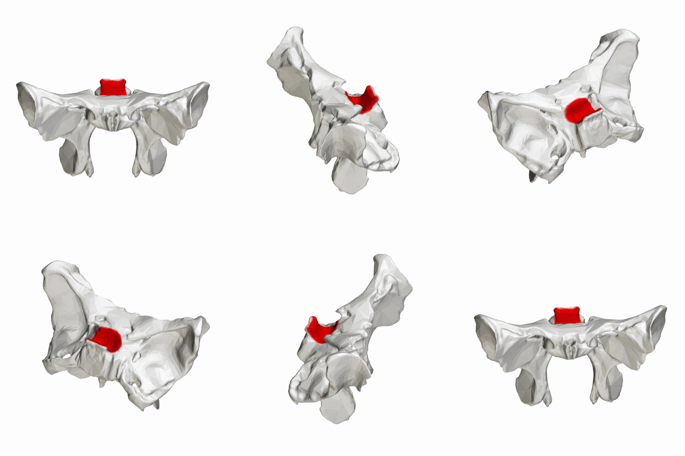
The sella turcica of the sphenoid bone

One of the features of the sphenoid bone is called the "sella turcica" or "Turkish saddle" which it apparently resembles because it has 4 horns (see animation on the left).

And finally, my favorite cocktail trivia piece from today: do you know where the temple on the side of the head gets its name? There are quite a few well-reasoned urban myths regarding prayer and whatnot, but these are totally off-base. As a clue, the temple shares it's name with the temporal bone over which it located.

Do you have your guess?

Both features are named for the passage of time, specifically because the hair around the temple is usually the first to turn gray as we age.

WILTIMS #49: Mnemonics

Only a pair of anatomy tests today, so I don't have anything new to share. I will, however, leave you with a couple mnemonics.

First, for remembering the carpal bones of the hand:
Some Lovers Try Positions That They Can't Handle
Scaphoid, Lunate, Triquetrum, Pisiform, Trapezium, Trapezoid, Capitate, and Hamate

Second, for remembering the order of the anatomic entities that pass under the flexor retinaculum of the ankle:
Tom, Dick And Very Nervous Harry
Tibialis posterior tendon, flexor Digitorum longus tendon, posterior tibial Artery, posterior tibial Vein, tibial Nerve, and flexor Hallucis longus tendon

Saturday, October 12, 2013

WILTIMS #48: Groans

Today we only had one one hour lecture, so I don't have much to report. The topic was clinical correlations of lower extremity anatomy, which lead to many a groan as we watched every major traumatic athletic injury in recent memory.

It's been amusing how each lecturer has treated these grotesque images. Our course director used them sparingly and with an impressive degree of nonchalance. The radiologist warned us in advance and even said that the squeamish could cover their eyes until we had passed the slide. The surgeon, on the other hand, displayed his dozens of examples with enthusiasm and theatricality. As he was speaking today I happened to be sitting next to the course director. He groaned and winced just like the rest of us.
The reactions of teammates to Kevin Ware's broken leg (you've been warned)
TIL: The singular form of the word meninges is meninx. The meninges are the three layers of connective tissue (the dura mater, arachnoid mater, and pia mater) that cover the brain and spinal cord. And since they are almost exclusively described together, we never use the singular. Meningitis is the inflammation of the meninges, which is particularly dangerous because it can damage the enveloped nervous tissue. The meninges also have poor blood supply,  limiting both the body's immune response and the effectiveness of medications.